. 2019 Jul 11;10(35):8171–8178. doi: 10.1039/c9sc00193j

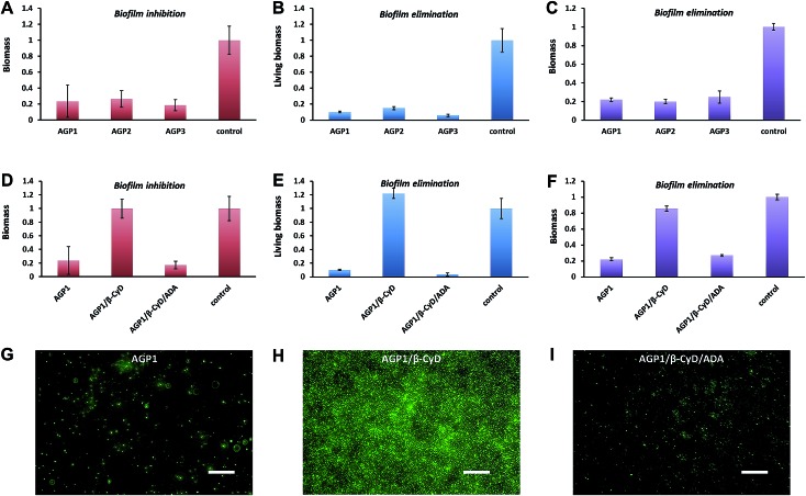
Fig. 6. (A and D) The biomass of the formed S. aureus biofilms in the presence of 0.6 mM AGP1, AGP2, AGP3, AGP1/β-CyD or AGP1/β-CyD/ADA for 24 h at 37 °C as quantified by crystal violet assay. (B and E) The living biomass of the preformed S. aureus biofilms after treatment with 0.6 mM AGP1, AGP2, AGP3, AGP1/β-CyD or AGP1/β-CyD/ADA for 24 h at 37 °C as quantified by XTT assay. (C and F) The biomass of the preformed S. aureus biofilms after treatment with 0.6 mM AGP1, AGP2, AGP3, AGP1/β-CyD or AGP1/β-CyD/ADA for 24 h at 37 °C as quantified by crystal violet assay. (G, H and I) Fluorescence microscopy imaging of the formed S. aureus biofilms in the presence of 0.6 mM AGP1, AGP1/β-CyD or AGP1/β-CyD/ADA for 24 h at 37 °C after SYTO9-staining. The scale bars are 20 μm.

Fig. 6