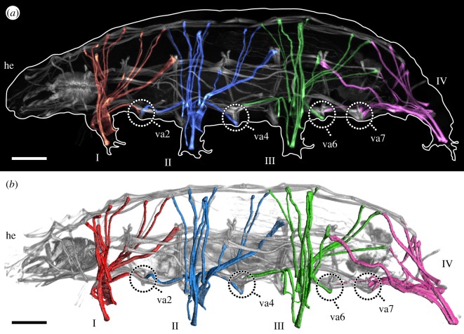
Figure 1.
Overview of the muscular system of the eutardigrade H. exemplaris. Colour-coding according to leg. Lateral view; anterior is left, dorsal is up in both images. Note that ventral attachment sites 1, 3 and 5 are located at the same level as legs I, II and III, respectively, and are therefore not labelled. (a) CLSM substack showing the muscular system of the left half of the animal, with leg muscles artificially coloured by leg. F-actin labelling. (b) Three-dimensional reconstruction based on the CLSM dataset shown in (a). he, head; I–IV, legs I–IV; va2–va7, ventral attachment sites 2–7. Scale bars, 20 µm.

