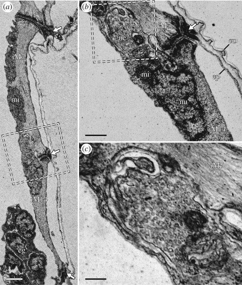
Figure 11.
Fine structure of iM14, with details of its proximal attachment site. STEM-in-SEM images. Sagittal sections; anterior is left, dorsal is up. White arrows indicate muscle attachment sites. (a) Overview of the posterior region of leg I showing iM14 and the distal region of iM7. (b) Close-up of the region outlined in (a) showing the iM14 proximal attachment site. A neuromuscular junction is adjacent to the attachment site. (c) Close-up of the region outlined in (b) showing the neuromuscular junction, which is full of synaptic vesicles. cg, claw gland; cu, cuticle; ep, epidermis; mi, mitochondria; ne, neuromuscular junction; nu nucleus. Scale bars, 1 µm (a); 500 nm (b); 200 nm (c).

