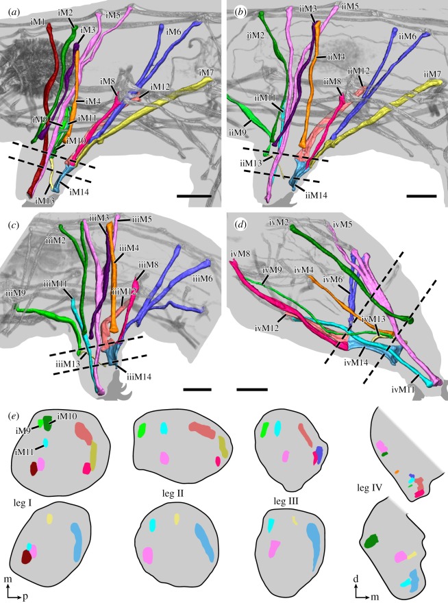
Figure 4.
Leg muscles of the tardigrade H. exemplaris. Three-dimensional reconstructions of legs I–IV (a–d, respectively) based on CLSM datasets of F-actin labelling. Dotted lines indicate planes of cross-sections shown in (e). Colour-coding according to hypothesized serial homologues. Lateral view; anterior is left, dorsal is up. (a) Muscle iM1 (red) does not share a serial homologue in any other leg. (c) Muscle iiiM2 (green) shows a branching morphology, unique to leg III. Muscle iiiM6 (blue) consists of three strands, with the posteriormost strand possibly representing a functional analogue of iM7 and iiM7. (d) Leg IV is rotated backwards relative to legs I–III so that ivM14 occupies an anteroventral position within the leg. Muscle ivM5 (pink) consists of three strands. (e) Schematic diagrams illustrating cross-sections of legs I–IV. Virtual sectioning planes are indicated by dotted lines in (a–d); top row corresponds to upper plane, bottom row to lower plane. Note how muscles of legs I–III are concentrated either at the anterior or posterior region of the leg, with muscle M13 spanning the two regions. Muscle M14 occupies the majority of the distal posterior region of legs I–III. Cross-sections not drawn to scale. Orientations in (e) for legs I–III (left) or leg IV (right). Orientation legends: d, dorsal; m, medial; p, posterior. Scale bars, 10 µm (a–d).

