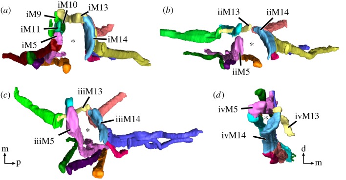
Figure 5.
Leg muscles of the tardigrade H. exemplaris from the distal perspective. Colour-coding according to hypothesized serial homologies. Three-dimensional reconstructions of legs I–IV (a–d, respectively) based on CLSM datasets of F-actin labelling. Note how muscles are arranged around the periphery of each leg—primarily in the anterior and posterior regions of each leg—with a large space in the middle (asterisk). The M13 muscles are the only muscles that cross between the anterior and posterior regions. (d) The muscles of leg IV are laterally compressed, presenting a smaller empty space between the muscles. Orientation in (c) for (a–c). Orientation legends: d, dorsal; m, medial; p, posterior.

