Figure 6.

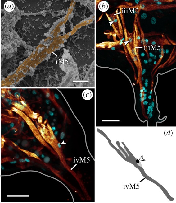
Branched morphologies in leg muscles. Lateral views; anterior is left, dorsal is up in all images. (a) False-coloured scanning electron micrograph of a bisected specimen showing iM5. (b,c) CLSM substacks; anti-myosin II immunolabelling (glow) and nuclear counterstain (cyan). (b) Leg III. The branched muscles iiiM2 and iiiM5 with their respective nuclei (arrowheads) located near the point where the two strands of each muscle split. (c,d) Muscle ivM5 and its three strands. Note how its nucleus (arrowhead) is located below the point where the three strands split. Scale bars, 3 µm (a); 10 µm (b,c).
