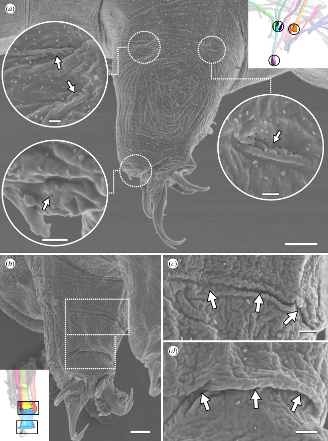
Figure 8.
Lateral and posterior leg muscle attachment sites seen on the external cuticle. Scanning electron micrographs. Insets (in a,b) show three-dimensional reconstructions based on F-actin labelling of the same perspectives with corresponding regions outlined. (a) Lateral and distal anterior muscle attachment sites of leg III. Each attachment site (outlined and shown as a close-up) is recognizable by what appear to be tiny holes in the cuticle (arrows) and, in some cases, a ridge or fold. Lateral view; anterior is left, dorsal is up. (b–d) Attachment sites of iM14. (c,d) shows close-ups of the upper and lower outlined areas in (b), respectively. Posterior view of leg I; medial is right, dorsal is up. Note the wide, thick folds of the cuticle at both the proximal (c) and distal (d) iM14 attachment sites. Scale bars, 5 µm (a); 500 nm (insets in a); 3 µm (b); 1 µm (c,d).

