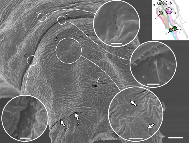
Figure 9.
Posterior end of the body showing the attachment sites associated with leg IV. Scanning electron micrographs. Inset shows three-dimensional reconstruction based on F-actin labelling of the same perspective with corresponding regions outlined. Posterodorsal view; anterior is upper left. Muscle attachment sites are recognizable by what appear to be tiny holes in the cuticle. Outlined regions of interest (a–d) are shown in close-ups. Distal attachment sites (e,f) are located under the fold in the leg in this specimen. cm, attachment site of the cloacal muscles. Scale bars, 4 µm (overview); 500 nm (close-ups of a–c); 2 µm (close-up of d).

