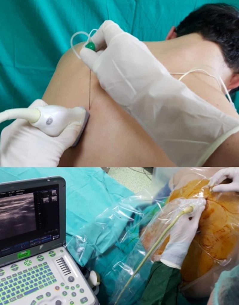
Figure 2. The position of the patient, ultrasound probe, and needle during the performance of an erector spinae plane block.
The ultrasound probe is placed 2-3 cm lateral to the spinous processes in the longitudinal plane. The needle is inserted from the cephalad aspect of the probe and advanced in the caudal direction within the plane of the ultrasound beam.

