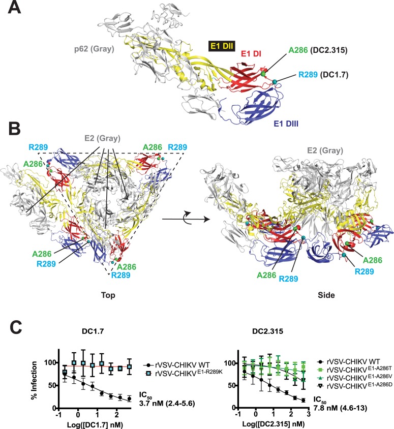
Fig 5. Viral escape studies with E1-targeting mAbs.
Location of A286 and R289 on the p62-E1 X-ray structure (A, PDB ID: 3N40) [9] or on the E1/E2 cryoEM heterohexamer (B, PDB ID: 3J2W) [11]. For clarity, p62 or E2 subunits are colored gray whereas E1 domains DI, DII, and DIII colored as per Fig 1. In panel B, a complete prefusion E1/E2 hexameric spike (outlined with dotted line) is illustrated, along with an E1/E2 heterodimer from an adjacent spike, to depict relative orientation within adjacent spikes. (C) Neutralization studies with WT VSV-CHIKV and viral escape mutants. Data are pooled from two experiments, each performed in duplicate or triplicate (points represent mean ± SD).

