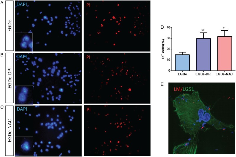
Figure 4.

PI staining analysis of U251 cells after invasion by L. monocytogenes. (A) DAPI and PI staining of U251 cells after invasion by EGDe without treatment. (B) DAPI and PI staining of U251 cells after invasion by 2.0 μM DPI-treated EGDe. (C) DAPI and PI staining of U251 cells after invasion by 1.0 mM DPI-treated EGDe. (D) Statistical analysis of apoptotic cells in (A–C). Apoptosis ratio was calculated by dividing the number of PI-positive cells with that of DAPI-positive cells. Student t-test was used for statistical analysis, *P < 0.05, **P < 0.01. (E) Immunostaining of L. monocytogenes. As no permeation was taken, only extracellular bacteria can be labeled (Red), intracellular bacteria and cell nucleus were labeled with DAPI (Blue), U251 cells were labeled with Acti-stain 488(Green).
