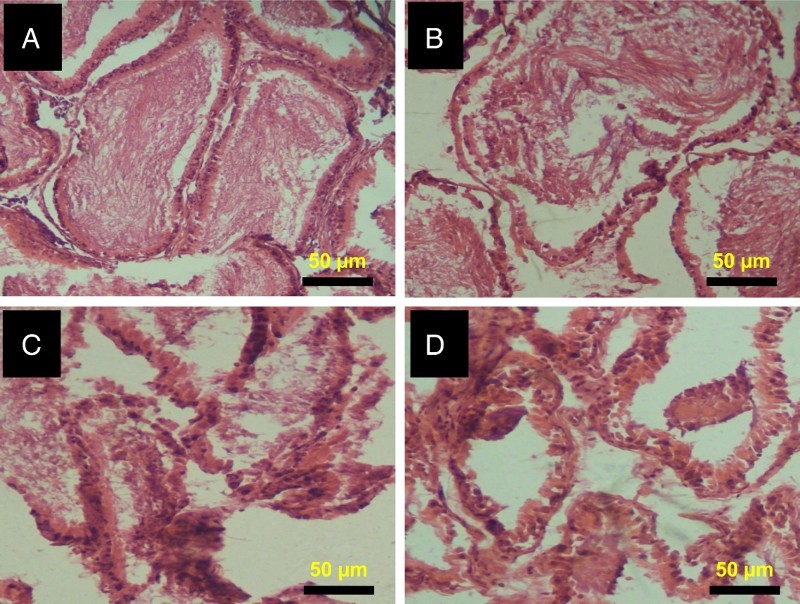
Figure 6.

Histopathology of epididymis from control and 2,5-HD-treated rats. Representative photomicrographs of epididymis from control (A) and 0.25% 2,5-HD (B) showed normal morphology. Epididymis from 0.5% 2,5-HD (C) and 1% 2,5-HD (D) groups showed progressive degeneration of epididymis characterized with severe erosion of the epididymal lining and reduced epithelia layer integrity.
