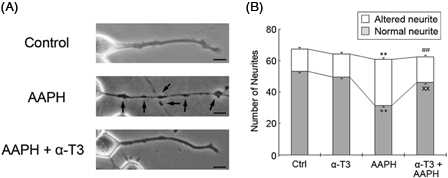
Figure 4.

α-Tocotrienol prevents AAPH-induced neurite degeneration. Neuro2a cells were treated with α-tocotrienol (5 µM) in the presence or absence of 1 mM AAPH. After 24 hours, the cells were fixed with 4% PFA in PBS. Photomicrographs of the cells were collected and analyzed on a personal computer. The scale bar represents 10 µm. Arrows indicate bead formation on the degenerating neurites of neuro2a cells (A). The results of quantitative analysis of neurite degeneration are shown (B). Each column represents the mean of three independent experiments. At least three wells were examined per experiment. Data were analyzed using the Student's t-test. ++P < 0.01 between normal control neurites and neurites of neurons treated with 1 mM AAPH. xxP < 0.01 between normal neurites of neurons treated with 5 µM α-tocotrienol in the presence or absence of 1 mM AAPH. **P < 0.01 between altered neurites of control neurons and those of neurons treated with 1 mM AAPH. ##P < 0.01 between altered neurites at 5 µM α-tocotrienol in the presence or absence of 1 mM AAPH.
