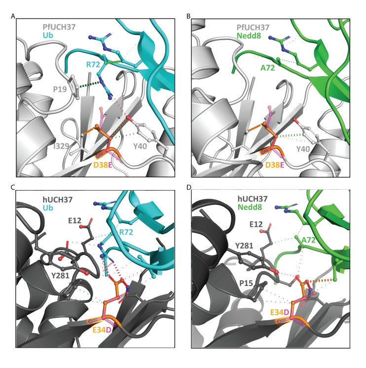
Fig 3. Structural models showing the effects of the mutations on the ubiquitin and Nedd8 binding pocket of UCH37 enzymes.
Ribbon representations of PfUCH37 (light grey) and HsUCH37 (dark grey) in complex with A) and C) human ubiquitin (cyan) and B) and D) HsNedd8 (green). The wild type UCH37 residues are shown in orange, and the mutant in magenta. Polar interactions are shown in orange, hydrogen bonds in red and hydrophobic interactions in green.

