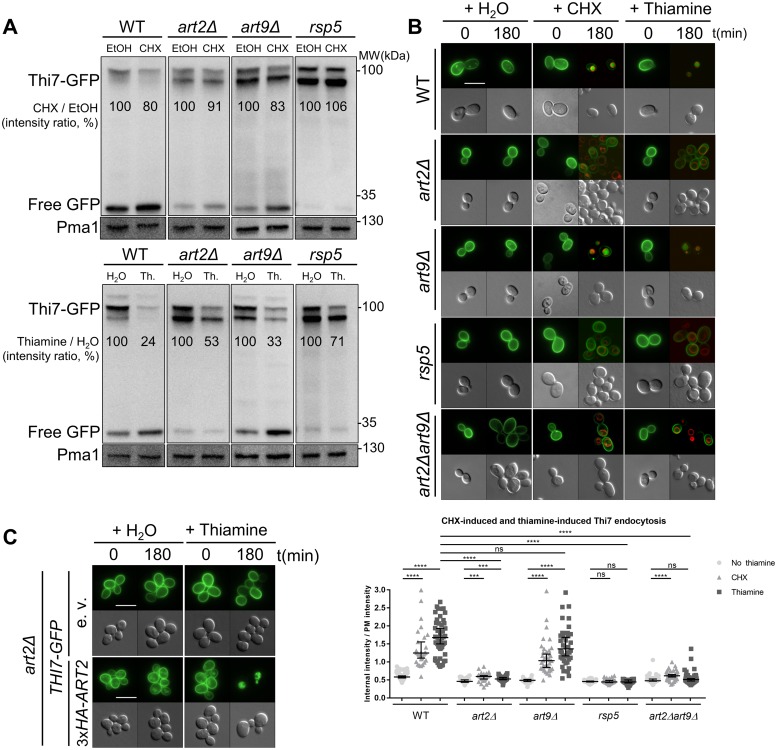
Fig 1. Art2 and Rsp5 are required for CHX- and thiamine-induced Thi7 endocytosis.
(A) The WT, art2Δ, art9Δ, and rsp5 strains expressing THI7-GFP under their endogenous promoter were grown in thiamine-free selective medium up to early-log phase and harvested after 120 min with CHX (final concentration; 50 μg/ml) or EtOH (top panel) and with thiamine (“Th.,” final concentration: 100 μM) or water (H2O) (bottom panel). Total protein extracts were immunoblotted with anti-GFP and anti-Pma1 as a loading control. Free GFP is the results of the degradation of Thi7-GFP in the vacuole. Thi7-GFP band intensity is normalized by the intensity of the Pma1-corresponding band. Representative of 3 experiments. (B) Localization of Thi7-GFP in the WT, art2Δ, art9Δ, rsp5, and art2Δart9Δ strains after addition of thiamine or CHX or mock-treated with water. The vacuolar membrane is stained with FM4–64. Quantification shows the ratio of internal-over-PM fluorescence intensity as described in Materials and methods section (n > 30 cells) (****p < 0.0001; ***p < 0.001; **p < 0.01; *p < 0.05). (C) Expression of 3xHA-ART2 restores thiamine-induced endocytosis of Thi7-GFP in the art2Δ background. THI7-GFP and either 3xHA-ART2 or its corresponding e.v. are expressed in the art2Δ strain, grown in thiamine-free selective medium, and complemented with thiamine (final concentration: 100 μM). Scale bar represents 5 μm. The numerical data are included in S1 Data. CHX, cycloheximide; EtOH, ethanol; e.v., empty vector; GFP, green fluorescent protein; ns, nonsignificant; PM, plasma membrane; Pma1, plasma membrane ATPase 1; WT, wild type.

