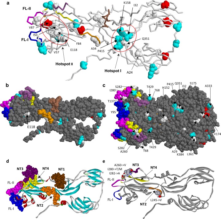
Fig 7. Location of amino acid substitutions in 3D structure of E1 glycoprotein.
a. E1 monomer with all the identified substitutions mapped. The models of mutations were based on the coordinates from PDB entry 4adg. Mutational hotspots are shown encircled by a red line, the hotspot I including the residues I32, D34, K158, Q351, P415 and the hotspot II including the residues I50, V57, F84, V87, E118. The mutation A24->V is common in all iVDRV strains studied. The mutation F84->L was found in 5 of 6 strains and is located in the vicinity of the fusion loops. The color codes used: the variant residues found both in RVs and RVi (white spacefill), only in RVs (cyan spacefill), only in RVi (red spacefill); neutralizing epitopes NT1 (residues 225–235, brown), NT2 (245–251, orange), NT3 (260–266, violet) and NT4 (274–285, yellow); fusion loops (FL-I, residues 89–98, blue; FL-II, residues 130–138, magenta). b, c. Mutated amino acid residues on the surface of E1 monomer (b) and trimer (c) in iVDRVs (solvent accessibility >0.5, probe size 1.4A). d. The NT epitopes in E1 monomer. e. Possible escape mutations in the E1 neutralizing epitopes NT2, NT3 and NT4.

