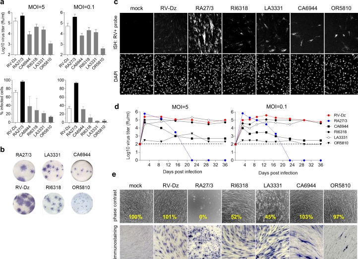
Fig 8. iVDRV growth properties.
a. Virus yields and percentages of RV-positive cells after infection of WI-38 with iVDRV, wtRV and RA27/3 strains at MOI = 5 (2 dpi) and MOI = 0.1 (3 dpi). Virus titers in the media were determined by titration on Vero cells, the number of infected cells was estimated by immunostaining for E1 protein. Data are presented as a mean +/- s.d. (n = 3, each experiment was performed in duplicate). b. Foci of infection of iVDRV isolates on Vero cells in comparison to wt and vaccine foci revealed by immunostaining for E1 at 6 dpi. c. Representative images from two independent experiments showing the results of the RNA-FISH for positive-strand RV RNA in WI-38 mock infected or infected with RA27/3 or RV-Dz at MOI = 5 (2 dpi). Nuclei were counterstained with DAPI. d. iVDRV persistence in WI-38. Growth curves of the indicated strains were shown for MOI = 5 and 0.1. Media were collected every 3–6 days and the extracellular viruses were titered on Vero cells. The representative results of two independent experiments each done in duplicate are shown. Limit of the assay detection (1x10² ffu/ml) is depicted by the dashed line. e. Phase contrast images of mock infected or infected (MOI = 5) cells at 36 dpi. Note cytopathic effects of RI6318 and LA3331 and the lack of live cells in RA27/3-infected wells. The adherent cells from one of two duplicate wells at 36 dpi were counted; the percentage of remaining adherent cells in each well was calculated relative to the mock infected well (yellow text). The cells in the second duplicate well were immunostained for E1.

