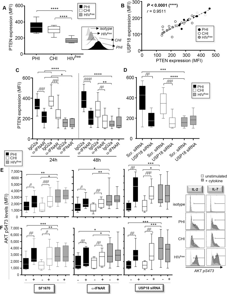
Fig 2. High USP18 expression in Mem from PHI and CHI impairs AKT activation.
(A) Ex vivo PTEN expression levels in Mem from PHI, CHI and HIVfree subjects (MFI) (n = 10). Representative histograms including isotype control are also shown above. (B) Correlations between USP18 expression and PTEN expression in Mem for all subjects (n = 30). (C) PTEN expression levels in Mem that have been treated for 24 or 48 hours with α-IFNAR or its respective isotype control (n = 10). (D) Expression of PTEN in Mem that have been transfected with siRNA specific for USP18 or with scrambled siRNA (n = 10). (E, F) Levels of AKT pS473 following 15 minutes of IL-2 (E) or IL-7 (F) stimulation in Mem that have been pre-treated 48 hours with SF1670 (left), α-IFNAR or its respective isotype control (middle), or transfected or not for 48 hours with specific USP18 siRNA (right) (n = 10). Representative histograms for AKT p473 expression in cytokine-stimulated Mem for all groups of subjects including isotype control are also shown on the right side. The error bars indicate standard deviations from the means. β, symbol used for paired t test (comparison between treated Mem and control). *, symbol used for Mann-Whitney test (comparison between study groups).

