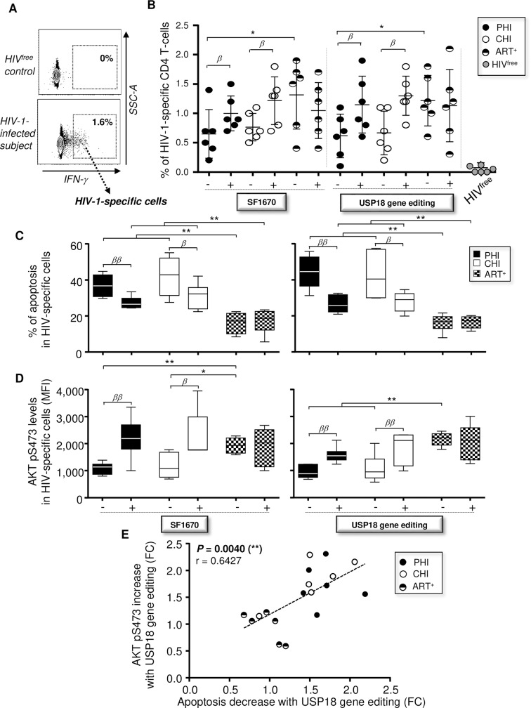
Fig 7. Interfering with USP18 reduces apoptosis of HIV-1-specific CD4 T-cells in an AKT-dependent manner.
(A) Gating strategy for HIV-1-specific CD4 T-cells following Gag stimulation for 18 hours. HIV-1-specific clones were determined by IFN-γ expression. (B) Percentages of HIV-1-specific CD4 T-cells (on total CD4) in CHI, PHI, ART+ and HIVfree subjects after Gag stimulation in the presence or absence of SF1670. Percentages of HIV-1-specific cells were also determined in culture after 18 hours of Gag stimulation when CD4 T-cells have been pre-transduced or not for 48 hours with LVUSP18 KO. HIVfree donors were included as negative control for HIV-1 stimulation (n = 6). (C,D) Levels of apoptosis assessed by Annexin-V staining (C) and AKT pS473 expression (D) in HIV-1-specific CD4 T-cells at 18 hours post-stimulation in the presence of absence of SF1670 (left) or CRISPR/Cas9 mediated USP18 gene editing (right) (n = 6). (E) Correlation between the reductions of apoptosis and increases of AKT pS473 levels in HIV-1-specific CD4 T-cells after USP18 gene editing (FC, fold change; n = 18). The error bars indicate standard deviations from the means. β, symbol used for paired t test (comparison between treated Mem and control). *, symbol used for Mann-Whitney test (comparison between study groups).

