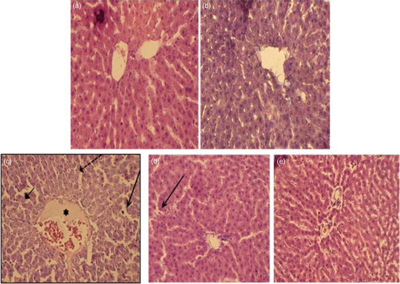
Figure 9.

Paraffin sections stained by H&E for histopathological examination of liver tissues of rats as follows: (A) control rats; (B) rats fed with berberine (50 mg/kg, i.g.); (C) rats treated with lead (500 mg Pb/l in the drinking water); (D) rats treated with lead (500 mg Pb/l in the drinking water) and fed with berberine (50 mg/kg, i.g.) and (E) rats treated with lead (500 mg Pb/l in the drinking water) and fed with silymarin (200 mg/kg, i.g.). The long arrow indicates leukocyte infiltration. The short arrow indicates pyknotic nucleus. The dashed arrow indicates hepatic cell necrosis. The asterisk indicates the distended portal vein.
