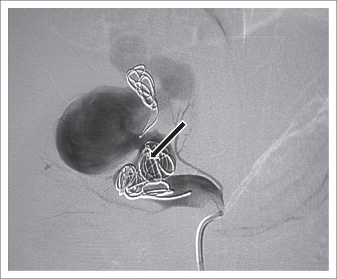
FIGURE 4.
Angiogram after the first-coil embolisation demonstrating the first micro coil that migrated to the venous side because of high flow. The ectatic segment of the feeding artery was packed with coils but despite that there was still filling of the arteriovenous malformation because of high flow.

