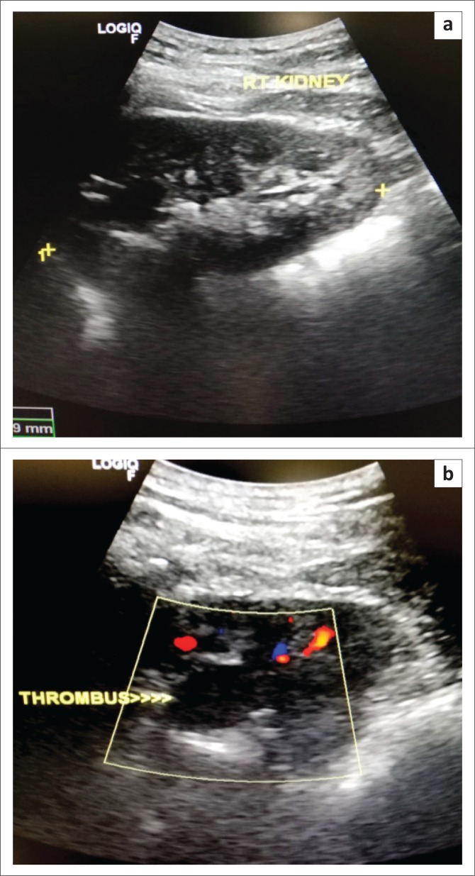
FIGURE 6.
Post-embolisation Ultrasound and Doppler demonstrating a thrombus in the feeding artery and absence of venous flow 4 weeks after the procedure: (a) Post-embolisation Ultrasound of the right kidney showing a thrombus in the feeding artery at the right upper pole. (b) Post-embolisation Colour Doppler demonstrating absence of venous flow.

