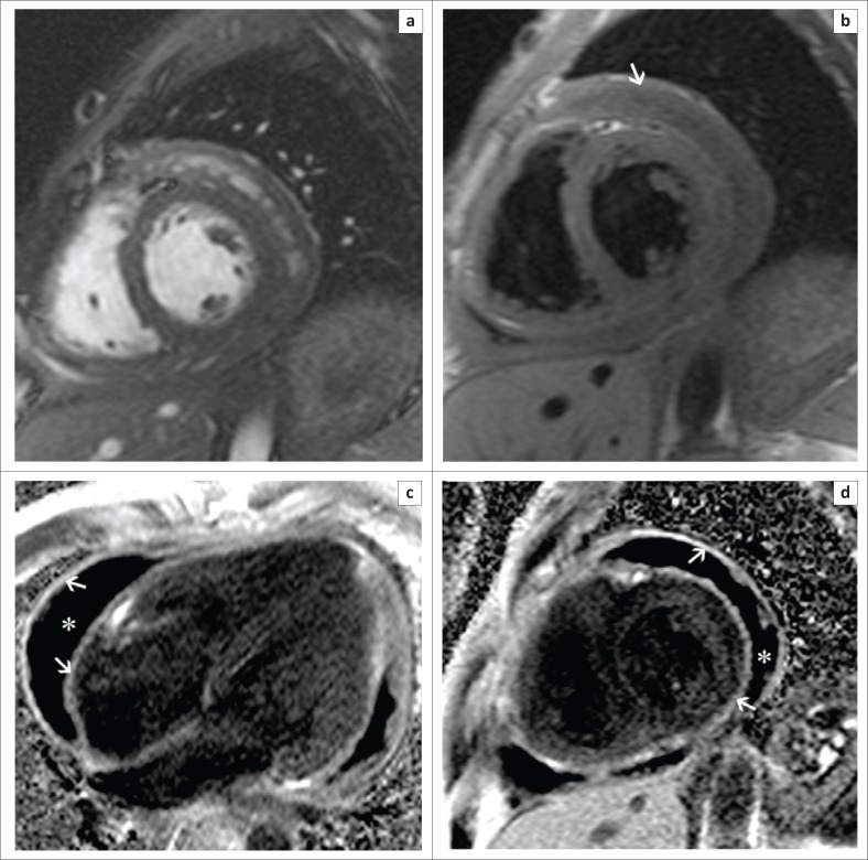
FIGURE 8.
Short-axis SSFP (a) and T1-weighted (b) images demonstrate the presence of a large pericardial effusion (white arrow) and marked thickening of the visceral and parietal pericardium. Four-chamber (c) and short-axis (d) late gadolinium images show intense enhancement of the thickened visceral and parietal pericardium (white arrows) and the large hypointense pericardial effusion (white asterisk).

