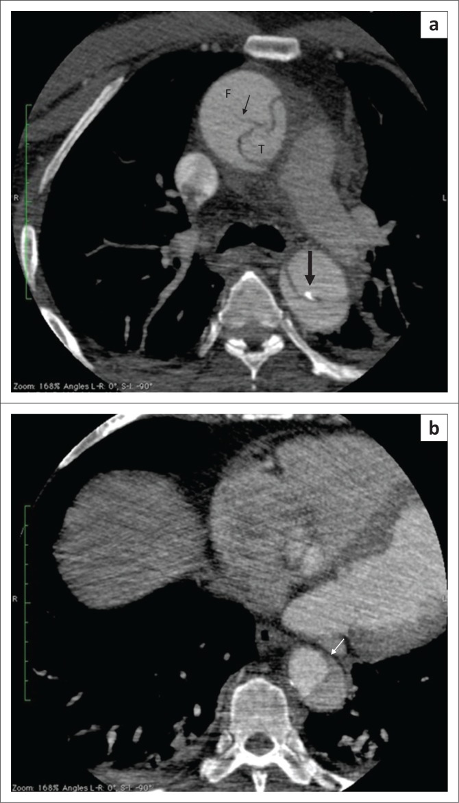
FIGURE 3.
(a) Contrast-enhanced axial computed tomography (CT) scan showing intimal calcifications around the true lumen (thick arrow) and linear scattered hypodense areas within the false lumen termed the Cobweb sign (thin arrow), representing the remnants of media. The calibre of the true lumen (T) appears smaller than the false (F). (b) Axial contrast-enhanced CT scan showing a beak sign (arrow) which is a wedge of haematoma at the distal end of false lumen forming an acute angle with the vessel wall.

