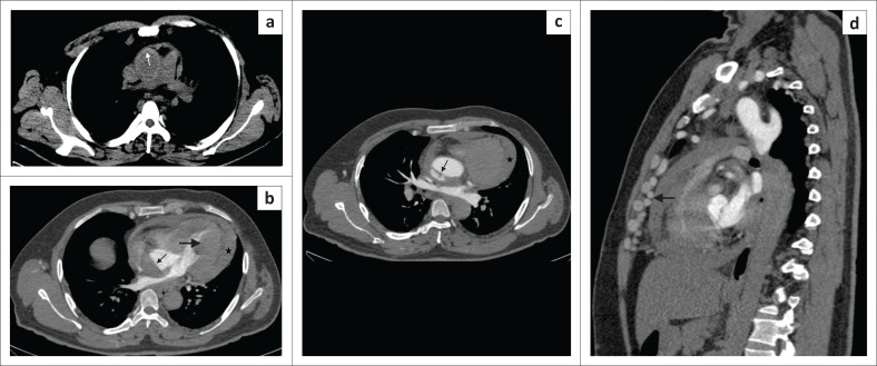
FIGURE 8.
Coarctation of the aorta with a type A intramural haematoma (IMH) and aortic dissection. (a) Unenhanced axial computed tomography (CT) scan showing circumferential hyperdense thickening of the ascending aorta (arrow); (b) Contrast-enhanced axial CT showing crescentic non-enhancing hypodense area (thin arrow) representing IMH with haemopericardium (*) and concentric left ventricular hypertrophy (thick arrow); (c) Contrast-enhanced CT scan showing the displaced intimal flap (arrow) and haemopericardium (*). (d) Sudden narrowing of the aorta (arrow) distal to origin of the left subclavian artery on sagittal contrast-enhanced images. Multiple collaterals (thick arrow) along the anterior chest wall.

