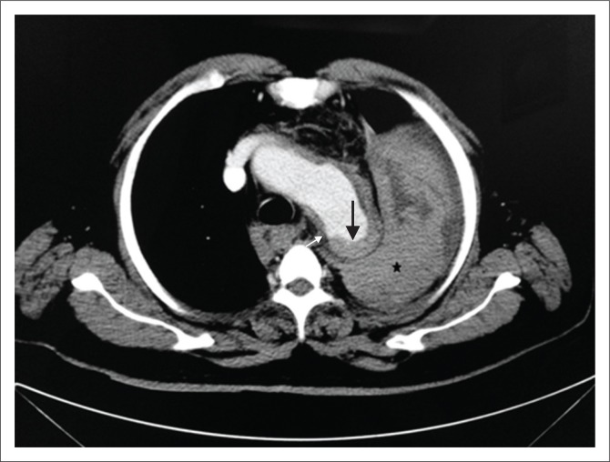
FIGURE 10.
Ruptured penetrating atherosclerotic ulcer (PAU) with contained haematoma. Contrast-enhanced computed tomography scan of thorax showing a ruptured PAU along the posterior aortic wall (white arrow) with adjacent intramural haematoma (black arrow) appearing as hyperdense aortic wall thickening and a contained haematoma in left pleural space (*). Findings were confirmed at surgery.

