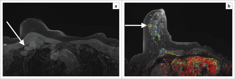
FIGURE 2.
Mammogram and breast ultrasound (not shown) did not reveal any pathology in either breast. Clinically, there were enlarged lymph nodes in right axilla and lymph node biopsy revealed metastatic adenocarcinoma. (a) Axial dynamic post-contrast MRI shows multiple enlarged, enhancing lymph nodes (white arrow). (b) A small, irregular enhancing mass is evident in the anterior half of the right breast. Dynamic post-contrast scan with kinetic colour overlay demonstrating mostly red (i.e. washout that is highly suspicious for malignancy). On kinetic overlay maps, red reflects washout, yellow plateau and blue persistent features on delayed post-contrast scans. Magnetic-resonance-imaging-guided biopsy was performed. High-grade invasive ductal carcinoma confirmed on histology.

