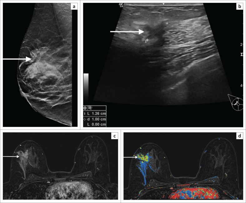
FIGURE 7.
(a) Mammogram (LCC) in a 42-year-old patient who had an area of architectural distortion in the upper half of the right breast (white arrow). (b) Ultrasound indicated a suspicious mass (white arrow) – irregular, anechoic mass with an echogenic halo. However, histology indicated a complex sclerosing lesion. This was felt to be discordant. (c) Post-contrast magnetic resonance imaging shows a spiculated solid mass (white arrow). (d) The colour overlay shows washout and plateau features. Repeat biopsy-confirmed invasive carcinoma.

