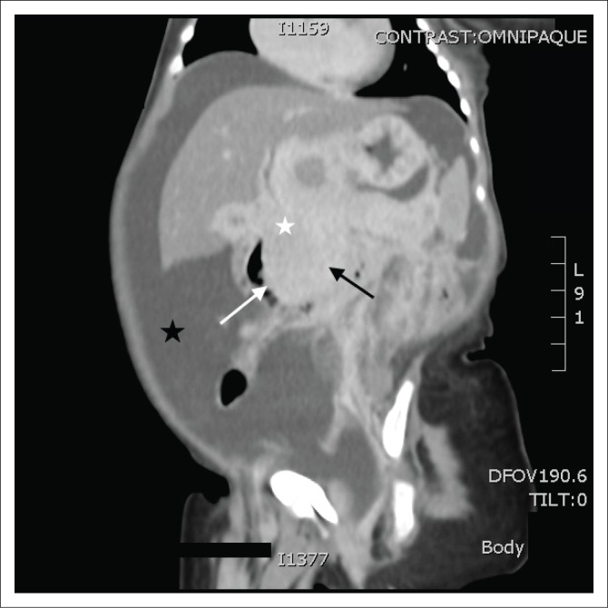
FIGURE 1.
Coronal post-contrast computed tomography image demonstrating an ill-defined, enhancing mass in the head of the pancreas (black arrow), with a poor plane of separation from the D2 segment of the duodenum (white arrow) and peripancreatic extension to the porta hepatis (white star). Secondary findings of ascites (black star).

