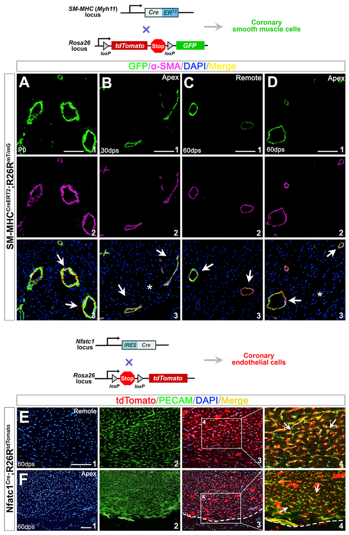
Figure 7. New Coronary SMCs and Endothelial Cells within the Ventricular Apex Are Derived from the Pre-existing Coronary Vessels.
(A) Immunostaining of SM-MHCCreERT2; R26RmT/mG hearts at P0 with tamoxifen induction during gestation.
(B–D) Hearts at 30 dps (B) and 60 dps (C and D) with apical resection at P1.
GFP+ cells (A1, B1, C1, and D1) are cells of SM-MHC lineage. (A2), (B2), (C2), and (D2) are antibody staining for α-SMA. (A3), (B3), (C3), and (D3) are overlays of SM-MHC lineage (GFP+) with DAPI and α-SMA. α-SMA+ SMCs in the injured apex are from SM-MHC lineage (arrows in B3 and D3). Asterisk indicates injured area.
(E and F) Regeneration of coronary endothelial after apical resection. Immunostaining of Nfatc1Cre/+;R26RtdTomato/+ hearts at 60 dps.
(E) Nfatc1 progeny give rise to coronary endothelium (arrows in E4).
(F) PECAM+ endothelial cells in the newly formed apex are from Nfatc1+ endocardial/endothelial lineage (arrows in F4).
(E1) and (F1) are DAPI staining; (E2) and (F2) are PECAM staining; (E3) and (F3) are overlays of Nfatc1 lineage (tdTomato+) with DAPI. (E4) and (F4) are high-magnification images of the areas outlined in (E3) and (F3). They are overlays of PECAM staining and Nfatc1 lineage (tdTomato+). Scale bar, 100 μm.

