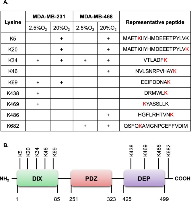
Figure 2.

Putative lysine residues acetylated on endogenous DVL-1 under different oxygen tension and their approximate location on DVL structural domain. (A) The table indicates putative lysine residues on DVL-1 that were found to be acetylated under different oxygen tension (2.5% O2 and 20% O2) in MDA-MB-231 and MDA-MB-468 cells along with their representative peptide as detected by liquid chromatography mass spectrometry (LC-MS/MS) analyses. (B) Approximate representation of position of acetylated lysine (K) residues on DVL-1 conserved domain is shown. NH2 represents N-terminal; followed by amino-terminal DIX domain, a central PDZ, and a carboxyl-terminal DEP domain, and C representing C-terminal.
