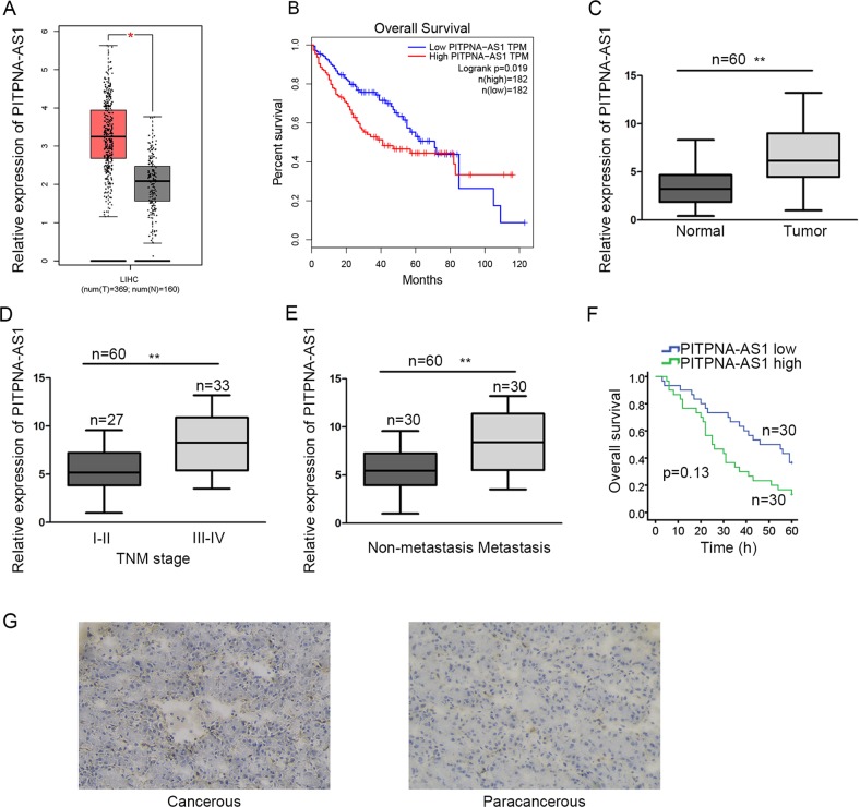
Fig. 1. PITPNA-AS1 was heightened in hepatocellular carcinoma tissues.
a The expression pattern of PITPNA-AS1 in TCGA liver cancer patient samples was obtained from GEPIA. b Surviving curve of TCGA liver cancer patients was generated and downloaded from GEPIA in accordance with the median of PITPNA-AS1 expression in tumor samples. c–e qRT-PCR analysis of PITPNA-AS1 expression in tumor and normal tissues (c), tissues in different TNM stages (d), and tissues with or without metastasis (e). f Kaplan–Meier method was utilized to evaluate the survival of 60 HCC patients with high or low level of PITPNA-AS1. g Expression pattern of PITPNA-AS1 in cancerous or paracancerous tissues collected from 60 HCC patients was examined with ISH. *P < 0.05, **P < 0.01 indicated statistically significant differences. PITPNA-AS1 phosphatidylinositol transfer protein alpha antisense RNA 1, HCC hepatocellular carcinoma, GEPIA gene expression profiling interactive analysis, TCGA the cancer genome atlas, qRT-PCR quantitative real time polymerase chain reaction, TNM tumor-node metastasis, ISH in situ hybridization

