
Figure 2.
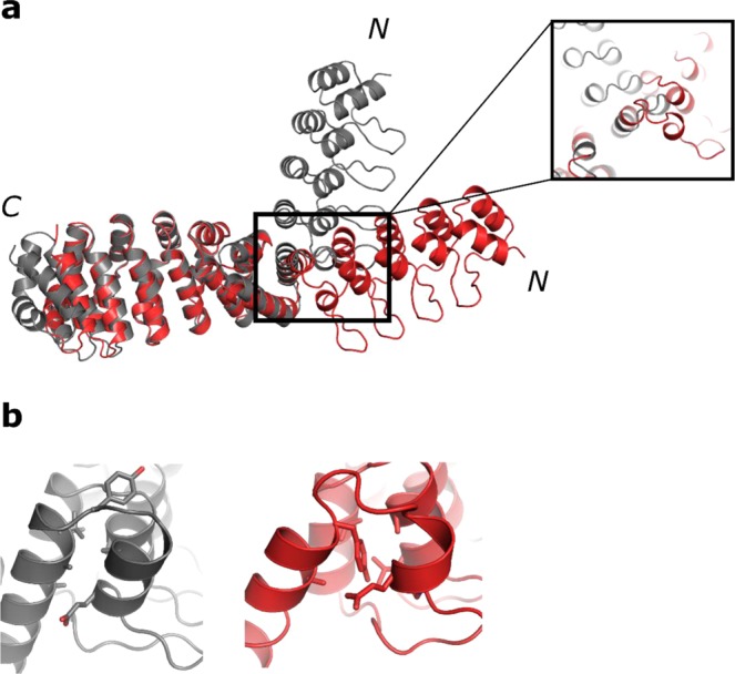
Comparison of model (grey) and crystal structure (red) of the fusion to helix 2 of the dArmRP. (a) Superposition of model (grey) and crystal structure (red, 1.6 Å resolution) of an N-terminal DARPin-dArmRP fusion. N- and C-termini of the proteins are marked. The fusion of DARPin D12 was made to H3 of a dArmRP with four internal repeats, with a shared helix length of 5 amino acids. The close-up view shows the distortion of the first helix of the DARPin. (b) Detailed view of the changed interface between model and experimental structure. The left picture is showing the model, the right one the structure in which Tyr150 inserts into the interface between the shared helix and the DARPin.