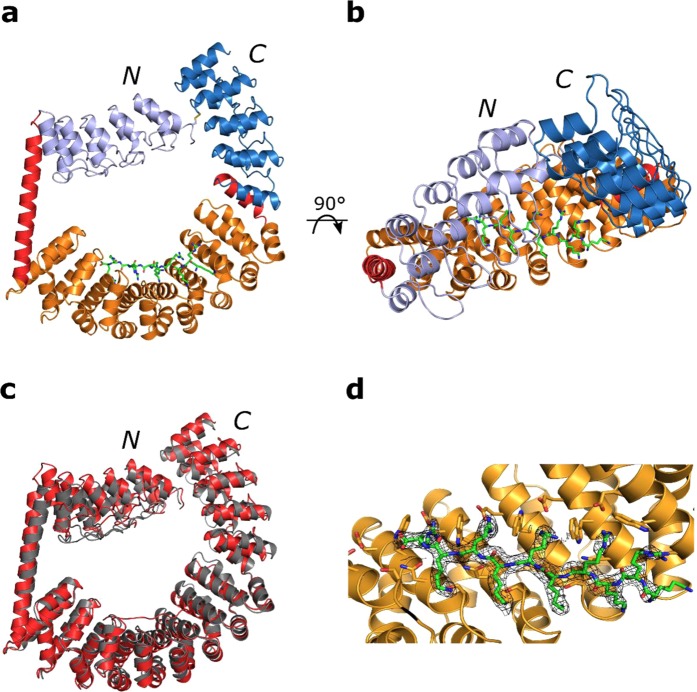
Figure 4.
Crystal structure of the ring-like construct showing a fully shielded binding surface at 2.4 Å. N- and C-termini of the proteins are marked. (a) Asymmetric unit with the DARPins coloured in light blue (N-terminal DARPin) and marine (C-terminal DARPin), the shared helices in red, the dArmRP in orange and the peptide in green. (b) Top-view highlighting the shielded binding surface (an overview of the crystal packing is shown in Fig. 5). (c) Overlay of the designed model in grey with the crystal structure in red. (d) Close-up view of the binding surface showing the regular peptide binding. 2mFo-DFc electron density of the peptide contoured at 1 σ.

