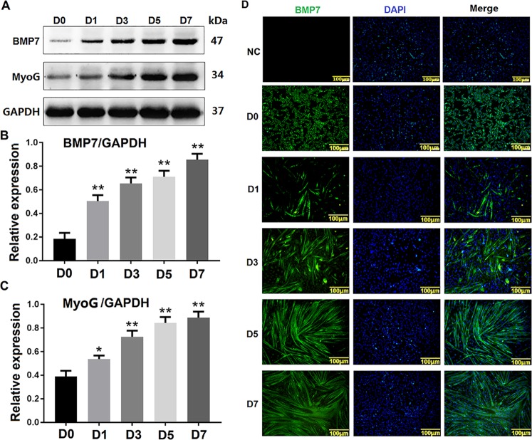
Fig. 4. Expression pattern of BMP7 during the differentiation of C2C12 cells.
a shows the western blotting results of BMP7 expression during the differentiation of C2C12 cells at 0, 1, 3, 5, and 7 days (D0, D1, D3, D5, D7). b, c shows the greyscale scans of BMP7 and MyoG from A. d shows the immunofluorescence results of BMP7 at different stages of C2C12 cell differentiation, NC is a negative control for BMP7 antibody. D0, D1, D3, D5, and D7 represented C2C12 cells induced to differentiate at 0, 1, 3, 5, and 7 days, respectively. The green fluorescent signal is BMP7 protein, while the blue fluorescent signal is the nucleus. The scale bar in D is 100 μm. **P values < 0.01 and *P values < 0.05 were considered as significant

