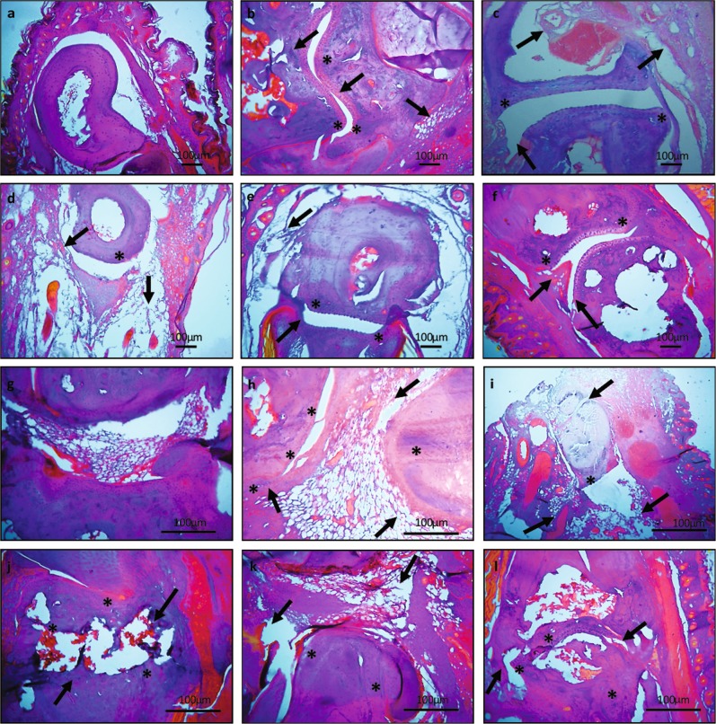
Fig. 3. H&E staining of ankle and tarsal joints.
a Normal mice tarsal joint revealed intact cartilage and no signs of bone erosion. b Arthritic tarsal joint exhibited joint modification, continuous bone erosion and cellular infiltration. c Hydroxychloroquine treated mice depicted diminished cellular infiltration and pannus formation along with progressive bone erosion of tarsal joint. d The tarsal joint of leflunomide treated group had modification in joint architecture but reduced cellular infiltration. e There was less cellular infiltration and diminished joint modifications after aqueous extract administration. f The ethyl acetate extract treated mice had preserved joint structure along with controlled cellular infiltration. g Normal ankle joint. h Arthritic ankle joint exhibited cellular infiltration joint modification and continuous cartilage degradation. i Hydroxychloroquine treated mice depicted progressive cartilage degradation. j The ankle joint of leflunomide treated group had joint modification along with cartilage distortion. k The cartilage and joint architecture was preserved along with less cellular infiltration in ankle joint of aqueous extract treated mice. l However, the ethyl acetate extract treated mice had preserved joint architecture and less cartilage distortion but still there were morphological changes. Arrows indicate joint modification, pannus formation, cartilage, and bone erosion whereas * indicate cellular infiltration. Original magnifications ×20 and ×40. Scale bars are shown below

