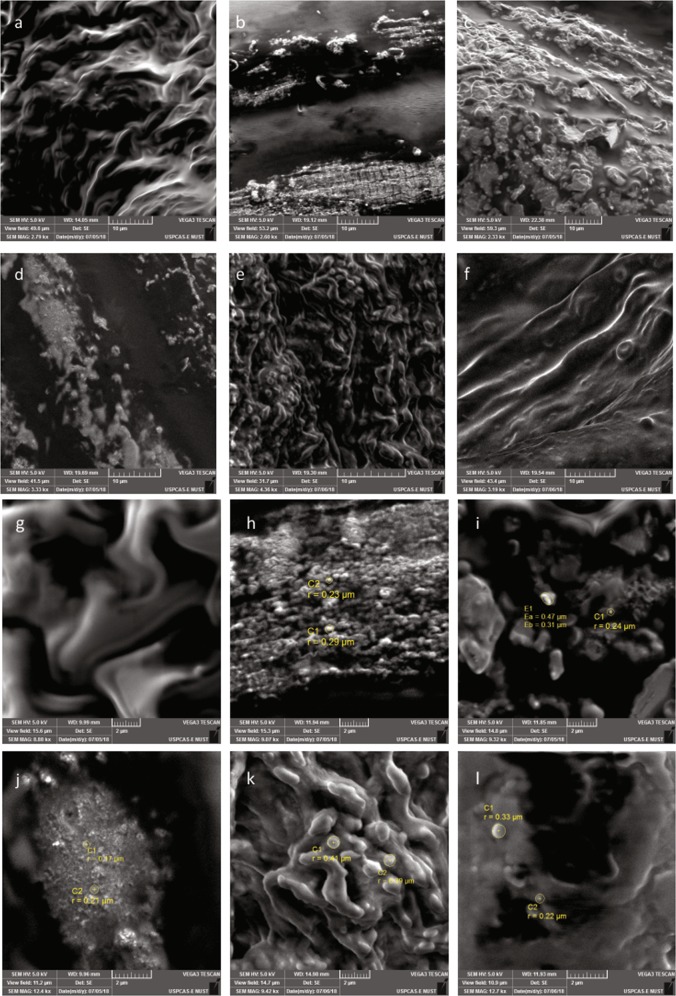
Fig. 5. Scanning electron micrographs showing cellular morphology of skeletal muscles along with signs of apoptosis and autophagy.
a Normal skeletal muscles had no signs of cellular degeneration. b Whereas the arthritic muscles had signs of aggressive muscular degeneration. c Hydroxychloroquine treated mice explicitly demonstrated signs of apoptosis in skeletal muscles. d The leflunomide treated group had signs of apoptosis but there were few indications of cell engulfing. e There are evident signs of apoptosis in aqueous extract treated mice. f The ethyl acetate extract treated mice had morphology near to normal with evident signs of restored balance of autophagy and apoptosis. g Normal skeletal muscles indicated no signs of aggressive apoptosis and autophagy. h Arthritic mice depicted signs of aggressive autophagy and engulfing of cellular debris due to muscular disintegration. i Hydroxychloroquine treated mice depicted progressive apoptosis as inferred from blebs observed in the micrograph. j The leflunomide treated group indicated signs of apoptotic bleb formation but the autophagy seemed diminished. k The skeletal muscles of aqueous extract treated mice had explicit signs of apoptosis and clearance of the extracellular matrix through cell engulfing. l However, the ethyl acetate treated mice exhibited signs of apoptosis with clearance through autophagy. Scale bars are provided below

