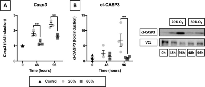
Figure 4.
Activation of caspase-dependent apoptosis. Samples were obtained after slicing (0 h) and after 48 or 96 h of incubation at either 20 or 80% O2 (n = 3–4). Baseline mRNA expression of Casp3 (A) was examined via qPCR. Cleavage of CASP3 (B) was investigated by western blotting, using vinculin (VCL) as a loading control. Values represent individual experiments performed in triplicate and are accompanied with the arithmetic mean (horizontal line) ± standard error of the mean (error bars). (**p < 0.01).

