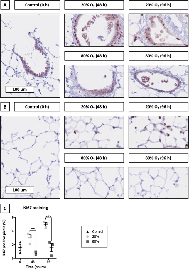
Figure 8.
Localization of proliferating cells. Samples were obtained after slicing (0 h) and after 48 or 96 h of incubation at either 20 or 80% O2 (n = 3). Ki67 stainings were performed to localize proliferating cells in the airways (A) and parenchyma (B). Subsequent algorithmic analysis revealed Ki67 positive pixels (C). Values represent individual experiments performed in triplicate and are accompanied with the arithmetic mean (horizontal line) ± standard error of the mean (error bars). (**p < 0.01 and ***p < 0.001).

