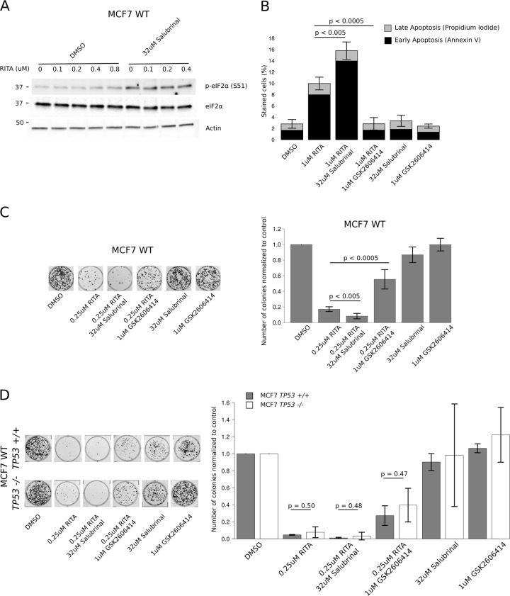
Fig. 5. Phosphorylation of eIF2α modulates RITA’s effect on apoptosis and colony formation.
a Western blot analysis using extracts from MCF7 WT cells treated with increasing concentrations of RITA in presence or absence of 32 µM salubrinal for 4 h. b FACS based quantification of Annexin V and propidium iodide staining to detect early and late apoptosis in MCF7 WT cells treated with vehicle (DMSO) or 1 µM RITA in presence or absence of 32 µM salubrinal or 1 µM GSK2606414 for 4 h (n = 3). c, d Crystal violet staining of WT (c), TP53+/+ or TP53−/− MCF7 cells (d) after treatment with vehicle (DMSO) or 1 µM RITA in presence or absence of 32 µM salubrinal or 1 µM GSK2606414 (4 h treatment followed by a 10 day expansion before staining). The pictures represent representative images and quantification was performed on n = 3 experiments. All bars represent the mean +/− SD

