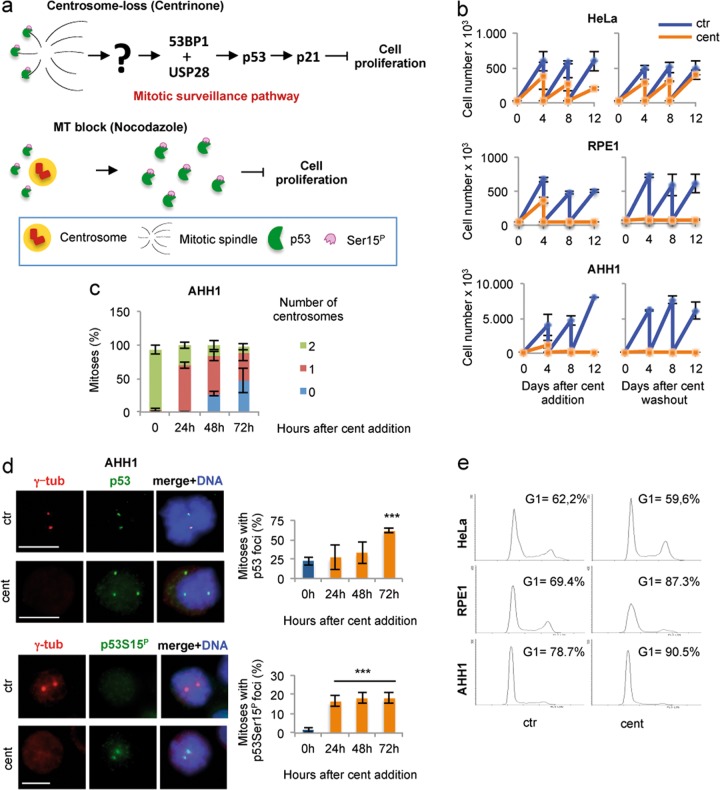
Fig. 6. p53-MCL as a centrosome-associated sensor.
a Upper panel: schematic representation of the mitotic surveillance pathway16 activated by centrosome-loss induced by centrinone treatment15. Lower panel: schematic representation of the accumulation of the p53Ser15P foci induced by inhibition of MT dynamics by nocodazole-treatment23. b Cell passaging assays were performed with the indicated cells after the addition of DMSO (ctr) or centrinone (cent) and after their washout. HeLa and RPE1 cells behaved as reported15. The nontransformed AHH1 LCL irreversibly arrested after centrinone treatment as well as RPE1 cells. c Time course analysis of centrinone-induced centrosome-loss in AHH1 mitotic cells. The percentage of mitoses with 0, 1, and 2 centrosomes has been calculated by analyzing at least 50 mitoses per time point. d Representative IF images of ctr and cent-treated AHH1 cells (48 h time point) obtained by double IF for γ-tubulin/p53 and γ-tubulin/phosho-p53Ser15 show that centrosome-loss associated with the formation of extra-centrosomal p53 foci that can be phosphorylated at Ser15. Histograms show the percentages of mitotic cells with p53 and p53Ser15P foci at the indicated time-points after centrinone addition. e The indicated cells, ctr or cent treated for 8 days, were analyzed for DNA content by flow cytometry. The relative cell-cycle profiles show that AHH1 cells accumulate in the G1 phase of the cell cycle. RPE1 and HeLa cells were used as controls and behaved similarly to previous report15

