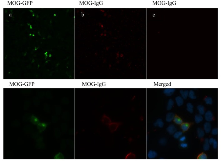
Figure 1.
MOG-IgG antibodies detected by cell-based assay (CBA). HEK-293A cells expressing human full-length MOG-GFP (green) (a) following addition of MOG-IgG-positive RON serum sample (b) and negative MS serum sample (c) (titer of 1:160). MOG-IgG antibodies were visualized using a Cy3-conjugated goat anti-human IgG antibody (red). Images are shown in 20× (upper panel) and 100× (lower panel) magnification.

