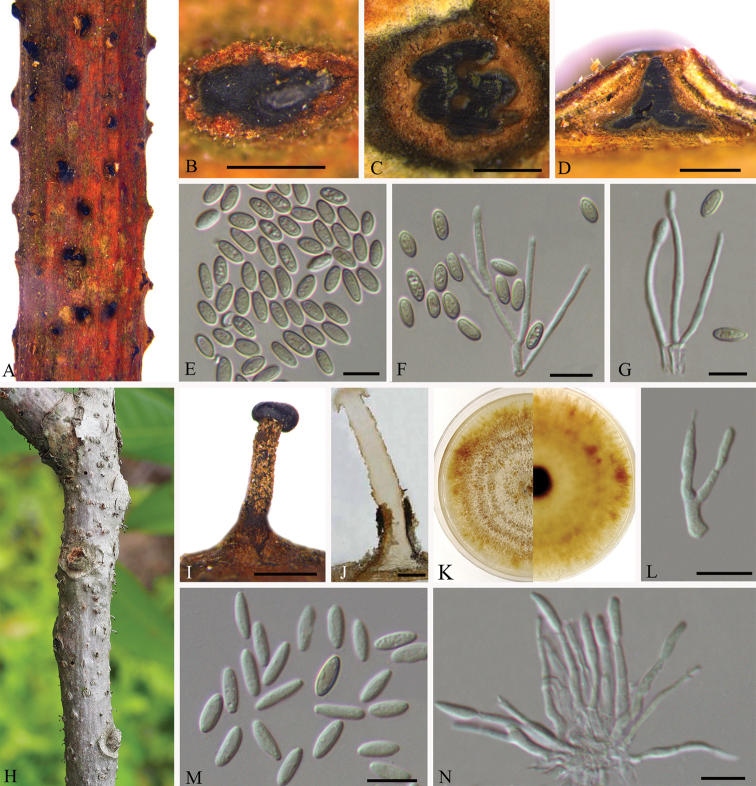
Figure 3.
Asexual morphology of Synnemasporella aculeans on Rhus chinensis (BJFC-S1740) A, B habit of pycnidia on twigs C transverse section of pycnidium D longitudinal section through pycnidium E conidia F, G conidiogenous cells and conidia H, I habit of synnemata on twigs J longitudinal section through synnema K the colony on PDA L, N conidiogenous cells bearing conidia M conidia. Scale bars: 500 μm (B–D, I, J); 10 μm (E–G, L–N).

