Fig. 1.

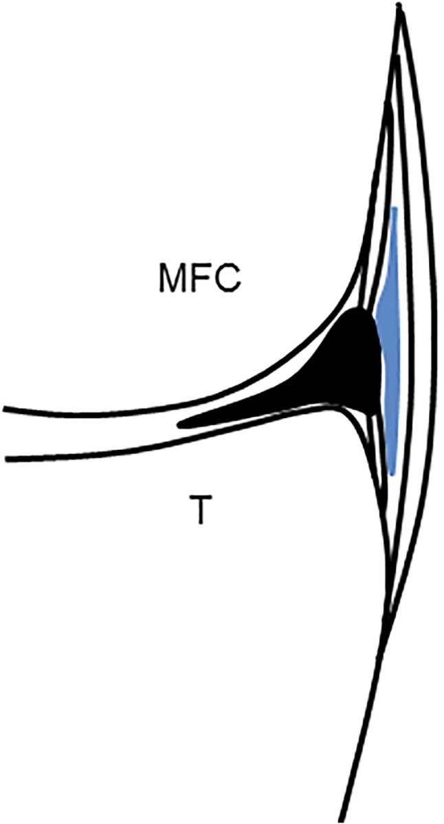
Schematic drawing shows a coronal section of the medial knee with a distended Voshell’s bursa (blue) between the superficial and the deep contingent of fibres of the MCL. MFC medial femoral condyle, T: tibia

Schematic drawing shows a coronal section of the medial knee with a distended Voshell’s bursa (blue) between the superficial and the deep contingent of fibres of the MCL. MFC medial femoral condyle, T: tibia