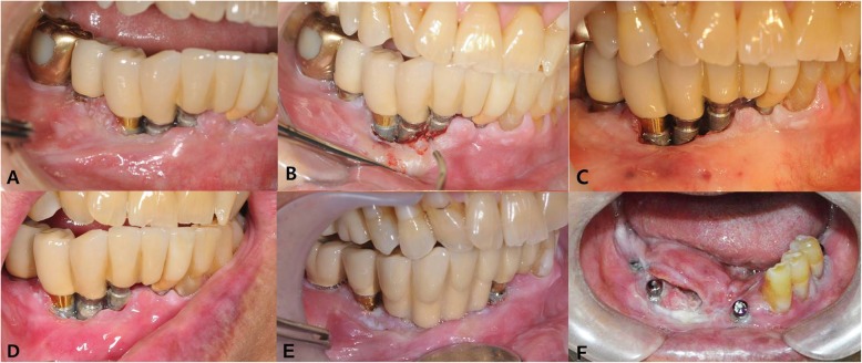
Fig. 1.
Clinical photos of the patient. a Initial visit; b peri-implantitis treatment with laser; c after 3-times laser treatment; d 5 months after initial visit; e 3 years after initial visit, the lesion was confirmed OSCC by incisional biopsy; f preoperative view show bulging tumor mass to lingual side

