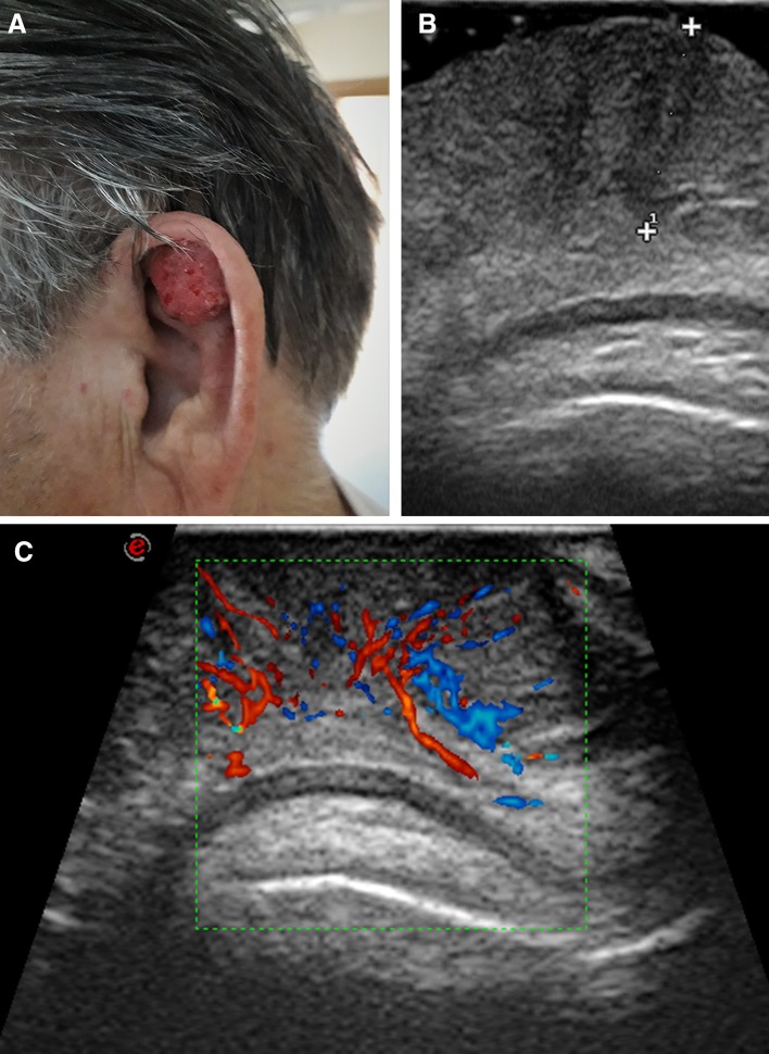
Fig. 4.
Ear scapha squamous cell carcinoma. Patient photograph (a). Ultrasound scan (b). The 7-mm thick lesion (calipers) is hypoechoic and relatively homogeneous. There is no infiltration of the cartilage. Color-Doppler scan (c). Discrete vascularization, with vessels mostly arranged vertically, starting from two deep vascular poles

