Figure 13.
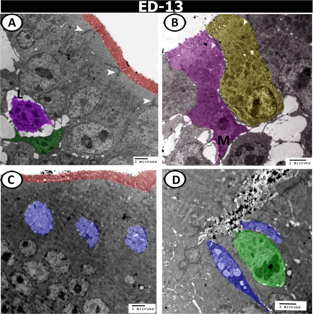
Digital coloured transmission electron microscopic (TEM) images of the proximal part of caeca on the 13th ED. (A,B) The epithelium consisted of simple columnar cells (yellow colour) with brush borders (red colour), and connected to the neighbouring cells by desmosomes (arrowheads). Note M-cell (M, pink colour), and the infiltration of the epithelium by dendritic cell (green) and lymphocytes (L, violet). (C,D) Goblet cells (blue colour), and rodlet cell (green colour) are distributed in the epithelium.

