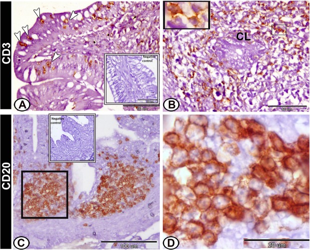
Figure 8.
Immunohistochemical expression pattern of CD3 and CD20 in the proximal part of caeca at hatching day. (A) Intraepithelial lymphocytes (arrowheads) expressed immunopostivity to CD3. (B) Lamina propria showed CD3-immunopositive T-lymphocytes (boxed areas) around the crypts of Lieberkuhn (CL). (C,D) CD20-positive B-lymphocytes were immunolocalised in the caecal tonsils (boxed area). Note, the negative control (boxed area) in A,C.

