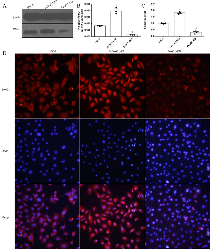
Supplemental Fig. 5.
Efficiency and specificity of 3aFoxO1-KI and FoxO1-KD in cultured HK-2 cells. (A) Representative western blot of FoxO1. (B) RT-PCR analysis of FoxO1 gene expression. (C) Quantitative analysis of the densitometry of FoxO1. (D) Immunofluorescence staining for FoxO1. *P < .05 vs. HK-2 cells; #P < .05 vs. 3aFoxO1-KI. P values were determined by one-way ANOVA analysis.

