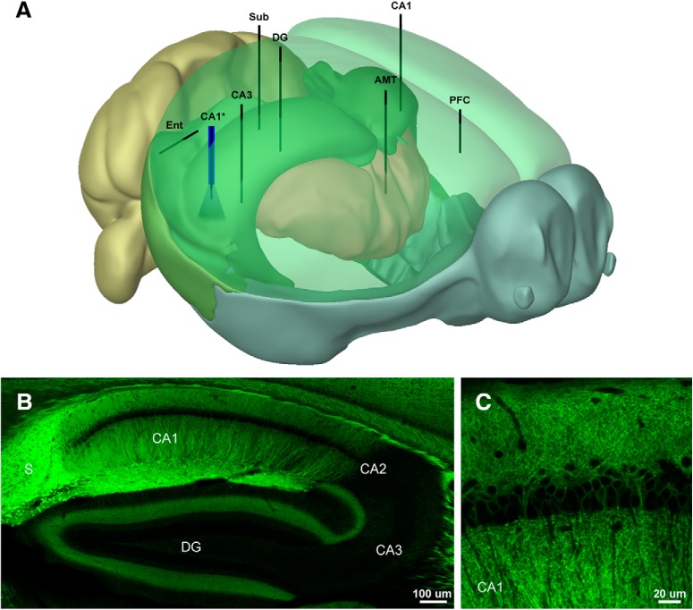
Figure 1.
Electrode placement and expression patterns in the Thy1-ChR2 (line 18) mouse. A, Schematic showing the placement of the electrode array and optrode. B, Distribution of ChR2-EYFP in the hippocampus of the Thy1 (line 18) mouse. Note high expression levels in CA1 and Sub; 10× tiled confocal image. C, ChR2-EYFP is expressed in deep pyramidal neurons in CA1 (Dobbins et al., 2018); 63× oil immersion confocal image. The 3D model was generated using Brain Explorer Software courtesy of the Allen Brain Institute (http://mouse.brain-map.org/static/brainexplorer).

